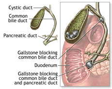
Gallstones that form in the gallbladder are the most common cause for blocked bile ducts. Additionally, bile duct stones can develop anywhere in the biliary tract where there is bile: within the liver, gallbladder and common bile duct. Gallstones and bile duct stones are usually comprised of cholesterol or bile salts — common components of bile — that have hardened into a stone. These stones can cause sudden pain when the cystic duct in the gallbladder or the common bile duct leading from the liver is blocked. Virginia Mason gastroenterologists treat this common problem in both adults and children with minimally invasive endoscopic technology. For more information or to schedule an appointment, call (206) 223-2319.
- Symptoms of Gallstones and Bile Duct Stones
- Diagnosing Gallstones and Bile Duct Stones
- Treating Gallstones and Bile Duct Stones
Symptoms of Gallstones and Bile Duct Stones
Gallstones can be miniscule in size or as large as a ping-pong ball. You may have one stone or develop many of them. Not all gallstones or bile stones cause symptoms. Some are discovered incidentally during imaging studies for other reasons.
The most common symptom is upper abdominal pain on the right side of the body, where the liver and gallbladder are situated. The pain may start suddenly and be intense. Or it may be a slow, dull pain or occur intermittently. The pain may shift from the abdominal area to the upper back or shoulder.
Prolonged blockage of a bile duct can cause a buildup of waste products in the biliary tract and in the bloodstream, leading to an infection called cholangitis. It also can prevent the release of bile into the small intestine to help digest food or cause a serious bacterial infection in the liver called ascending cholangitis.
A blocked bile duct may result in inflammation of the gallbladder, called cholecystitis. A gallstone or bile stone in the common bile duct may block the pancreatic duct, causing painful inflammation of the pancreas or pancreatitis.
If a stone completely blocks the ducts of the gallbladder, liver, common bile duct or pancreas, other symptoms may include:
- Nausea
- Fever
- Chills
- Yellow skin or eyes (from the build up of bilirubin, a waste product in blood)
- Dark urine
- Itching
- Fatigue
- Weight loss
- Night sweats
- Loss of appetite
- Greasy or light-colored stools
Patients who develop gallstones are at a slightly increased risk of developing gallbladder cancer, called cholangiocarcinoma. However, this is a rare disease and most people with gallstones do not go on to develop cancer.
Diagnosing Gallstones and Bile Duct Stones
Your gastroenterologist may suspect that you have gallstones or blockage of a bile duct based on your symptoms and results of a blood test showing high levels of bilirubin. Bilirubin is a waste product in blood caused from the normal breakdown of red blood cells.
Your gastroenterologist can diagnose and treat gallstones and bile duct stones at the same time with minimally invasive endoscopic technology. Common diagnostic tests and procedures for confirming the presence of stones include:
- BLOOD TESTS
In addition to a bilirubin test, your blood may be tested for the presence of elevated white blood cells used by the body to fight infection, and for abnormal levels of pancreatic and liver enzymes.
- ABDOMINAL ULTRASOUND
This non-invasive procedure uses sound waves rather than x-rays to produce images that can reveal gallstones and bile duct stones within the common bile duct. An ultrasound probe is passed over the abdomen and images are sent to a computer monitor. Abdominal ultrasound is commonly used in pregnant women.
- ABDOMINAL CT SCAN
A CT scan of the abdomen also can identify stones with the biliary tract and is a noninvasive procedure. During a CT scan images are shown on a computer monitor.
- ERCP
Endoscopic retrograde cholangiopancreatography, or ERCP, is a specialized endoscopic technique used to study the ducts of the gallbladder, pancreas and liver, and has the added benefit of being a therapeutic tool. ERCP has been used for more than 30 years. It is considered the standard modality for diagnosing and treating disorders of the biliary tract.
During this procedure, and after first receiving a mild sedative and an anesthetic to numb the throat, an endoscope containing a miniature camera is passed down your esophagus and into the biliary tract. When your gastroenterologist sees the biliary and pancreatic ducts, he or she then passes a catheter (a narrow plastic tube) containing a contrast dye through the endoscope. The dye is injected into the pancreatic and biliary ducts and X-rays are taken that are viewed on a computer monitor. The procedure takes 60 to 90 minutes and is performed in the Endoscopy Suite within Virginia Mason's Section of Gastroenterology and Hepatology.
Your gastroenterologist can treat a bile duct disorder at the same time it is being diagnosed by passing miniaturized instruments through the ERCP. Special preparations are required for this endoscopic procedure.
- ERCP WITH ENDOSCOPIC ULTRASOUND
Increasingly, gastroenterologists at Virginia Mason are using endoscopic ultrasound (EUS) in place of x-rays for better viewing of the bile and pancreatic ducts. During this procedure, an ultrasound probe is passed through the ERCP, which sends images to a computer monitor. Gastroenterologists can then treat disorders of the bile duct, including removal of gallstones and bile duct stones, with miniaturized instruments passed through the ERCP.
- MRCP
Magnetic resonance cholangiopancreatography is newer technology being employed at Virginia Mason. This noninvasive diagnostic procedure is performed using MRI technology that uses magnets and radio waves to produce computer images of the bile ducts. A contrast dye is injected first through the skin near the gallbladder to enhance the images. Patients are not required to undergo endoscopy preparation and they do not undergo sedation. MRCP is being used primarily in patients who may have failed or who are not good candidates for ERCP, in those who do not want to undergo an endoscopic procedure, and in individuals considered to be at low risk of having a pancreatic or bile duct disorder. While ERCP allows for therapeutic options with cholangioscopy, MRCP is a diagnostic tool only.
Virginia Mason also is involved in national clinical trials to determine the accuracy of MRCP in diagnosing disorders of the biliary tract.
Treating Gallstones and Bile Duct Stones
Gallstones and bile duct stones may be treated first with antibiotics to help control infection. They also can be treated at the time of diagnosis with miniaturized surgical instruments inserted through an ERCP. Alternatively, stones may be treated with medications that dissolve them, with lithotripsy that uses sound waves to break them up, or with surgery to remove the gallbladder.
- ENDOSCOPIC TECHNIQUES
When a stone has been identified on x-ray, ultrasound or MRI imaging as blocking a bile or pancreatic duct, it can be removed with miniaturized instruments inserted through the ERCP. These surgical instruments gently enlarge the ductal opening that then allows the stone to be removed.
- MEDICATIONS
Medications can be given that dissolve gallstones but they are not always effective and are not indicated in all cases. The most common medication is a bile salt (ursodiol) that slowly dissolves cholesterol within the stones. However, the stones can return when the medication is discontinued.
- EXTRACORPOREAL SHOCK WAVE LITHOTRIPSY
This treatment employs high-frequency sound waves to break up gallstones. Patients then take bile salt tablets, sometimes indefinitely, to dissolve the pieces and to ensure that the stones do not return. Only a minority of patients are candidates for this type of treatment, however. The best candidates have a single small stone. If an infection (cholangitis) or inflammation (cholecystitis) of the gallbladder is present, lithotripsy is not an option. Extracorporeal (meaning outside of the body) shock wave lithotripsy is performed by directing pulsating, high-intensity sound waves at the area where the stone is located, identified first by ultrasound. The procedure takes about 45 minutes and patients are usually lightly sedated before treatment.
- SURGERY
Surgery to remove the gallbladder, called cholecystectomy, is a common procedure in the United States for individuals with symptoms caused by gallstones. Virginia Mason was one of the first medical centers in the country to remove the gallbladder by the minimally invasive laparoscopic approach, called laparoscopic cholecystectomy.
This minimally invasive surgery for removing the gallbladder is one of the most common procedures performed at Virginia Mason and is, in fact, the preferred approach today for removal of the gallbladder. In cases in which a gallstone or bile stone has blocked a bile duct - a situation that can lead to infection or inflammation of organs within the biliary tract - surgeons will likely recommend removal of the gallbladder.
Laparoscopy
During laparoscopy, the surgeon makes several ¼ to ½ inch incisions in the abdomen. He or she then inserts miniaturized endoscopic and surgical instruments, and a small camera, through these "ports." Images from the camera are sent to a video monitor that allows the surgeon to "deflate" and then remove the gallbladder through one of the ports. Individuals return to their regular activities often within a few days.
Open surgery
Sometimes the surgeon must revert to an open surgical procedure during a scheduled laparoscopy to remove the gallbladder. These occurrences happen infrequently and are most often caused when the gallbladder is found to be infected or when the gallbladder lining is hardened, making it more difficult for the organ to be removed laparoscopically.
At other times, the surgeon may make the decision that the open surgical procedure is the best option for the patient based on the severity of the individual's gallbladder disease. Open surgery involves making a large incision in the abdomen and removing the gallbladder. Recovery time is longer, five to seven days in the hospital, and there is a longer return to daily activities: two to three weeks, for example.